Visual agnosia is a condition in which a person can see but is unable to recognize what it is they are seeing. The disruption occurs in the visual centers of the brain, where in the normal state not only the image itself is formed, but also the associative memory of it. That is, the disease is characterized by the inability to recognize sensory signals from the visual analyzer from a familiar object. In another way, visual agnosia is also called object or mental blindness.
It should be understood that the patient’s entire visual analyzer functions normally, there is no pathology in the refraction of light rays, their focusing on the retina, the formation of electrical impulses and their conduction to the brain. A comparison can be made to the blindness caused by optic atrophy. With it, some or all of the neurons responsible for conducting nerve impulses to the brain are lost. As a result, there is a violation of color and light perception, loss of areas of vision or complete blindness.
With visual agnosia, a person can copy a drawing, but cannot name what is depicted
In optical agnosia, signals from the eyes travel to the visual centers in the brain, but cannot be processed correctly. There is no synthesis of a single image from both eyes, or the association of pictures with an image or abstract concept is disrupted.
Therefore, the treatment of agnosia is closer to the field of neuropsychology.
What causes visual agnosia?
It is believed that visual agnosia occurs when areas of the cerebral cortex are damaged in its parietal and occipital-parietal lobes. It is here that information responsible for associations with objects is analyzed and synthesized. The parietal cortex stores all sensory information from the visual system, as well as tactile and spatial associations, sensory data from the skin about temperature, touch, and pain.
Visual images are formed in the brain, its damage causes optical agnosia
Lesions in these parts of the brain may be caused by:
- Stroke. Brain damage occurs due to disruption of its blood supply as a result of ischemia, thrombosis, arterial embolism, or hemorrhage. Nervous tissue, not receiving oxygen, begins to die, and gradually parts of the brain lose their functions.
- Neurological disorders and tumor processes. Visual agnosia occurs when there is massive damage to the occipital and parieto-occipital regions of the brain.
- Dementia. Neurodegenerative processes that occur in the brain with age lead to loss of cognitive functions and memory. The most common cause of dementia is Alzheimer's disease.
Other possible reasons:
- hereditary predisposition;
- meningeal infection;
- mechanical damage to the occipital lobes due to traumatic brain injury;
- carbon monoxide poisoning;
- recovery after long-term blindness.
Facial agnosia
Facial agnosia occurs when the right hemisphere of the brain (middle and posterior parts) is damaged. The degree of its severity varies: from impaired memory of faces in special experimental tasks (failure to recognize familiar faces or their photographs) to failure to recognize oneself in the mirror. In addition, a selective violation of either facial gnosis itself or memory of faces is possible.
What is specific about a “face” as a visual object compared to an object? The interpretation of a violation of facial gnosis as a deficiency in the holistic perception of an object is confirmed by data on the difficulties of playing chess that occur in patients with damage to the right hemisphere.
In addition, the perception of a face always contains the contribution of the individuality of the perceiver, who sees in the face something of his own, subjective, even if these are portraits of famous people. The specificity of the perceived face is both in its unique integrity, reflecting the individuality of the “sample”, and in the attitude of the perceiver to the original.
Agnosia is a disease characterized by a violation of certain types of perception, resulting from damage to the cerebral cortex and nearby subcortical structures.
When the projection (primary) parts of the cortex are disrupted, sensitivity disorders occur (hearing loss, impaired visual and pain functions). In cases where the secondary sections of the cerebral cortex are affected, the ability to perceive and process the information received is lost.
How to recognize
Most cases of this disease occur in older people who have experienced some degree of brain damage. But signs of visual agnosia can occur at any age.
To understand how everything looks from the first person, imagine the following: you see two circles and a crossbar between them and you cannot even imagine what it is and what it is used for. But a healthy person, when looking at this object, easily determines that these are glasses. He immediately “knows” that they fit on his face and help improve his vision.
Drawing of a person with simultaneous visual agnosia
Depending on where exactly the brain damage occurred in the occipital lobes, the characteristics of visual agnosia vary.
Optic nerve atrophy
- With object agnosia, there is no holistic perception of the object, but its individual parts can be identified. A person lists the characteristics of an object, but cannot find out what it is.
- Agnosia for faces (prosopagnosia) occurs when there is damage in the right hemisphere. A person cannot recognize people he knows by face or in a photo, and in severe cases, he cannot recognize himself.
- Simultaneous agnosia does not allow the patient to look at all objects that are in the field of vision. The violation manifests itself in misses and disorientation. In this state, a person cannot draw anything within a given area (for example, in a circle) or outline a figure.
- Visuospatial agnosia. This type is also called one-sided, or left-sided, agnosia and is characterized by “omission” of the left half of space and even one’s body. The lesion is located on the right side of the brain, and the synthesis of images from the two hemispheres into one does not occur. It manifests itself in the fact that a person “does not see” the left side of the text or drawing; when asked to draw an object, he also draws only its right side from memory.
- With agnosia for symbols (letters), the area of damage is determined at the border of the temporal and occipital lobes. The patient can copy letters and numbers, but cannot recognize and name them, and accordingly the reading skill is lost.
- Agnosia by color. It is characterized by the fact that, in principle, the patient can name the color if it is presented separately on a card, but when asked to name the color of a certain object (for example, strawberries), he experiences difficulty. Variants of the disorder: amnesia regarding the names of shades, inability to imagine a color (imagine it), cortical blindness (complete lack of color discrimination).
- Space agnosia with damage to the area in the upper occipital of the brain. There are varying degrees of disturbance in the perception of the coordinate system. The patient is not oriented in the concepts of left-right, up-down, cardinal directions, or the distance of objects, but normal recognition of objects is maintained. Spatial agnosia is often combined with impaired coordination of movement; accordingly, the ability to write and read suffers. The following types of visual agnosia regarding spatial perception are also possible: macropsia (objects seem larger) and micropsia (smaller than they really are), polymelia (perception of false limbs).
Tactile agnosia
Tactile agnosia is a violation of the recognition of shapes and objects by touch. Appears after damage to the parietal lobe of the right or left hemispheres. There are several types of agnosia of this nature:
- Object agnosia is a pathology in which the patient cannot determine the size, shape and material of an object, while he is able to determine all its signs;
- Tactile agnosia is the inability to recognize letters and numbers drawn on the patient’s hand;
- Finger agnosia is a pathology characterized by a violation of identifying the names of the fingers when touching them with the patient’s eyes closed;
- Somatoagnosia is the inability to identify parts of the body and their location in relation to each other.
Visual impairment
Sensory, relatively elementary types of visual impairment include a decrease (change) in color perception, photopsia (sensation of bright flashes, sparks), hemianopsia (loss of part of the visual field). With total destruction of the optic nerve, complete blindness of the corresponding eye occurs - amaurosis. With a pathological process surrounding the perimeter of the optic nerve, the effect of tubular vision may occur.
These disorders are associated with the pathology of individual parts of the visual analyzer. They are not directly related to higher visual functions, although they are their basis.
Gnostic visual disturbances are called visual agnosias . Munch first made this concept the subject of analysis (Munch, 1881). In animal experiments, he showed that removal of the occipital cortex in dogs leads to cortical blindness. The experimental dogs could bypass and jump over various obstacles, but at the same time they ignored a number of objects: they did not pay attention to their owner, a bowl of food, etc. After analyzing the results of the experiment, Munch came to the conclusion that the dogs developed “physical” blindness, caused by the loss of memory acquired during the perception of the image, which led to the loss of the dogs’ ability to recognize what they saw. Somewhat later, such disturbances in visual perception were described as an independent syndrome in people with local disorders of brain activity. S. Freud proposed to designate such violations by the term “agnosia.”
Visual agnosia was first described as a violation of many types of perception of visual information, while maintaining visual sensations. They are associated with distortions in the work of the secondary fields of the visual analyzer and the adjacent tertiary fields of the cerebral cortex (18th, 19th and adjacent tertiary fields). So, visual agnosia is a disorder of visual perception that occurs when the cortical structures of the posterior parts of the cerebral hemispheres are damaged and occurs with the relative preservation of elementary visual functions (visual acuity, visual fields, etc.) .
The variety of violations of visual-perceptual activity is determined by the partiality of its defect in relation to various types of visual material (real objects, colors, faces, etc.) and various levels of implementation of visual perception as a complex purposeful activity based on the actualization of past experience formed in ontogenesis (updating visual representations, holistic complex simultaneous perception of visual stimuli, the ability to consciously identify visually presented objects, etc.) (Fig. 6, 7,8). There may be pronounced overlaps between different types of visual agnosia, because neighboring areas of the brain that perform different functions in processing visual information can be damaged simultaneously with local brain lesions. The lack of a unified system for interpreting visual agnosia leads to different principles of their classification. When trying to generalize them, six types of visual agnosia are phenomenologically distinguished.
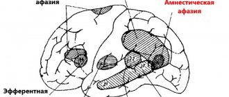
Object agnosia - occurs with damage to fields 18 and 19 (occipital and occipital-parietal zones, although there are cases of posterior temporal localization).
It can be characterized either as the absence of a recognition process, or as a violation of the integrity of the perception of an object with the possible identification of its individual features or parts. The inability to visually identify an object can be externally manifested as a listing of individual fragments of an object or its image (fragmentation), or
isolating only individual features of an object that are insufficient for its complete identification. Examples corresponding to these levels of manifestation of object agnosia can be the following: recognition of the image of “glasses” as a “bicycle”, because there are two circles; identification of a “key” as a “knife” or “spoon” based on the selected features “metal” and “long”. Thus, a person, while correctly assessing the individual elements of an object or its image, cannot understand its meaning.
Subject agnosia can have varying degrees of severity: from maximum to minimum. In severe cases (with bilateral damage to the 18th and 19th fields), visual recognition of individual real objects and their images is impaired. In moderately severe cases, patients do not recognize schematic, contour, inverted or superimposed images (in Poppelreiter tests), and difficulties arise in identifying objects with missing features or noisy objects. In milder cases, the time of tachistoscopic identification increases. Sometimes patients cannot imagine what a particular object looks like - a house, a tree, etc. With unilateral lesions of the occipital regions, differences in the structure of visual object agnosia can be distinguished. Damage to the left hemisphere is manifested to a greater extent by a violation of the perception of objects by the type of enumeration of individual details, while the pathological process in the right hemisphere leads to the actual absence of the act of identification.
Differential diagnostic signs of right hemisphere object agnosia are a slowdown in the process of identifying objects, a more accurate assessment by the patient of schematic images compared to realistic ones, as well as a narrowing of the scope of visual perception.
Facial agnosia (prosopagnosia) is associated with damage to the posterior parts of the right hemisphere (right infero-occipital region - the lower parts of the “wide visual sphere” - with a tendency to spread forward and to the inner surface of the temporal lobe). A number of publications indicate that bilateral lesions are mandatory. This is a selective gnostic defect; it can occur in the absence of objective and other agnosias. The degree of its severity varies: from impaired memory of faces in special experimental tasks, through failure to recognize familiar faces or their images (photos) to failure to recognize oneself in the mirror. In the presence of facial agnosia, for example, a woman with short hair may be mistaken for a man, etc. To recognize a person in such cases, auxiliary objects are used - voice, smell of perfume, features of gestures, gait. In addition, a selective violation of either facial gnosis itself or memory of faces is possible.
At the same time, such patients retain the ability to perform most mental tasks that require the use of visual information: for example, such people can read and name objects correctly.
It is assumed that the mechanism of this agnosia is a defect in the recognition of individualized features, because the categorical relationship to the image is preserved and faces are not confused with other objects. This type of visual agnosia can be designated as agnosia of individualized features while maintaining the categorical attitude to the image. Facial agnosia is one of the clearest illustrations of the simultaneous way of processing information characteristic of the right hemisphere. When the right hemisphere is damaged, the brain switches to a successive, sequential recognition strategy characteristic of the left hemisphere.
In foreign neuropsychology (I.M. Tonkonogiy, A. Pointe), two separate subtypes of facial agnosia are distinguished: apperceptive facial agnosia and associative facial agnosia .
Apperceptive facial agnosia matches the description above. It is a disorder of the earliest processes in the face perception system. In such patients, ideas about the desired face do not arise, and they are unable to make a choice from images of different faces. They also cannot distinguish between the age and gender characteristics of individuals. At the same time, they have some ability to compensate for the defect in the form of the ability to recognize people by voice, clothing, gait, hairstyle, etc.
Associative face agnosia is a disorder of the connection between the processes of face perception and the semantic information that is stored in memory about that person. Perceptual information usually has a certain meaning. Such a patient correctly identifies the differences between faces in two photographs offered to him and determines their age and gender characteristics. However, he cannot then identify the person, provide any information about his name, occupation, or the circumstances under which they last saw each other. With this agnosia, the face is perceived as such, but the person in it cannot identify what could become a distinctive feature.
Regarding the occurrence of facial agnosia, it was assumed that it is associated with brain lesions that are acquired during adulthood and (or) in childhood. But the results of recent studies (A.P. Bizyuk) suggest that a form of congenital facial agnosia also occurs, which in some cases is even hereditary.
Optical-spatial agnosia - occurs when the upper part of the “wide visual sphere” of the parieto-occipital region of the brain is damaged. The patient is poorly oriented in the spatial features of images or the reality surrounding him: he does not distinguish between “right and left”, does not understand geographical maps, watch dial indications, cannot copy a pose, cannot mentally rotate an object by 900 or 1800 (especially with the left hemisphere localization of the lesion lesions). When drawing complex geometric figures or faces, they cannot correctly arrange their fragments, are unable to copy the proposed pose, and do not recognize letters that have spatial characteristics. In more severe cases, orientation in the “top-bottom” coordinates is disrupted. In some cases, patients cannot localize objects in space; it becomes difficult to assess their distance and size, as a result of which even relatively simple everyday activities (dressing, eating) suffer. In some types of disorders, patients do not recognize well-known places. Some patients experience the phenomenon of visual depth agnosia, when they are unable to visually transfer three-dimensional objects onto a two-dimensional plane of a sheet, which requires taking into account perspective.
With unilateral lesions of the parietal-occipital regions on the right, the left part of the space is ignored, which seems to cease to exist (unilateral left neglect syndrome) (Fig. 9, 10). A possible reason for this defect is the impossibility of simultaneous synthesis of information coming from the left and right hemifields of vision. With bilateral parieto-occipital lesions, movement coordination disorders may be observed: a person’s hands begin to miss objects, which is a secondary praxis defect (apractoagnosia).
Letter agnosia - this type of agnosia occurs when the border between the occipital and temporal lobes of the left hemisphere (the lower part of the “wide visual sphere” on the left) is damaged. The patient, copying the letters correctly, cannot name them, because they are perceived simply as drawings without understanding their meaning. In this case, individual fragments of letters are not linked into a separate image of the sign. In some cases, the patient mixes up letters that are similar in spelling and experiences difficulties when moving from one font to another. In less crude cases, a person can read a word by tracing large letters with a finger. A similar structure of the disorder underlies color gnosis disorder.
Color agnosia - occurs with damage to the left occipital lobe and adjacent areas, but there is evidence of possible involvement of the left parietal-temporal region (to date, evidence has also been obtained of color perception disorders with damage to the right hemisphere of the brain). It is the least studied.
A distinction is made between color agnosia itself and color recognition disorders (color blindness), which, in particular, may be associated with damage to the retina or the lateral geniculate body. In the case of color agnosia, patients correctly distinguish individual, primary colors (for example, on cards), but are not able to associate a color with a specific object, or to sort objects by color. They also lose the ability to distinguish colors by shade and identify rarely encountered colors (terracotta, lilac, cyclamen, mustard), and their previously known names are forgotten. The patient, as a rule, cannot compare colors, match one color to another, rank colors by shade, or remember previously shown colors. This set of symptoms indicates the inclusion of the right hemisphere in the pathological process.
Simultaneous agnosia - occurs with bilateral or right-sided damage to the occipital-parietal parts of the brain. The cause of this type of agnosia is the “weakness” of visual cells, which are capable of only narrow local excitations. The essence of this phenomenon in its extreme expression is the impossibility of simultaneous perception of several visual objects or a situation in a complex. In this case, only one object is perceived or only one operational unit of visual information is processed, which is currently the object of human attention. For example, a person can perceive only individual parts of an object or its image, and no narrowing of the visual field is observed. In another case, patients correctly recognize individual objects and their details in the visual field or in pictures, but they cannot establish a connection between them and understand the meaning of the plot. This is combined with an inability to read words, but with preservation of the reading of individual letters. Simultaneous agnosia is not always clearly expressed. In less pronounced cases, only difficulties are observed in the simultaneous perception of a complex of elements with the loss of any details or fragments.
Quite often, simultaneous agnosia is accompanied by impaired eye movements and is considered within the framework of Balint's syndrome
, which is described as an independent pathology for bilateral lesions of the occipital lobes. It includes three symptoms:
– mental paralysis of gaze – the patient cannot look in a certain direction, but if an object accidentally comes into attention, the patient sees only it and does not perceive nearby objects;
– optical (oculomotor) ataxia – uncontrollability of gaze (inability to take an object under control of vision due to involuntary jumps of the eye, which is constantly in motion), which leads, for example, to the inability to read, because extraneous words constantly come into view;
– violation (narrowing) of visual attention.
However, this disorder should not be considered as a mandatory or the only mechanism for the formation of agnosia. The fact that the phenomenon of simultaneous agnosia is not directly related to gaze regulation disorders shows the independence of more complex levels of function organization, their relative independence from the state of simpler parts of the visual system.
In general, the nature of gnostic visual disturbances is quite heterogeneous. The nature of agnosia depends not only on the location of the lesion, but also on the degree of involvement of the commissural fibers connecting the hemispheres in the pathological process.
Constructive apraktoagnosia
Violation of voluntary movements and actions - aproxia. Constructive apraxia: apraktoagnosia, spatial apraxia. The main thing is the disintegration of complex spatial syntheses, i.e. performing movements and actions in conditions of spatial orientation - defects in locomotion (movement in space, the patient cannot find the way to the ward), poor orientation when moving on the ground, impairment when performing objective actions that require taking into account spatial relationships, apraxia of dressing (when putting on the thing is incorrectly positioned in relation to the body, difficulties when trying to get into the sleeve - usually occurs when the right hemisphere is damaged), there are disturbances in performing everyday activities (making the bed), difficulties in visual-constructive tests (drawing an object with a spatial location, copying a drawing , spatial recoding of the drawing, drawing objects relative to each other). The localization of damage is the inferior parietal areas of the left and right hemispheres of the brain.
Mechanism of development and manifestation
Simultaneous agnosia occurs due to right-sided or bilateral damage to the occipital-parietal region of the brain. The moment of perceiving information visually with this illness consists in the ability to process exclusively one operational unit of information, which at a given time is the object of attention.
For example, if you ask him to put a point in the center of a circle, he will not be able to cope with the task. In this case, you will need to perceive 3 objects simultaneously. We are talking about the boundaries of the circle, its center and the tip of a pencil or pen. In this case, the patient is able to see only one object out of the required three.
Quite often, simultaneous agnosia is accompanied by impaired reading skills. The patient is unable to read the entire word, although he is able to perceive each letter separately. At the same time, difficulties with their writing are not diagnosed.
Balint's syndrome is considered a serious manifestation of the pathology. With this disease, it is difficult for the patient to keep his gaze in only one direction or fix it on an object. Another manifestation of the disease is fragmented perception, which is characterized by seeing only part or detail of an object. For example, if you show a patient a table lamp and ask what he sees, the answer in the form of an ashtray will surprise many. The thing is that a person is only able to examine the lower part of an electrical device.
Agnosia and its varieties
Based on the type of incoming information, different types of agnosia are distinguished. All of them are united by one factor - the inability of the patient to perceive a certain type of information.
There are three main types of agnosia:
- visual;
- auditory;
- violation of tactile and spatial sensations.
Each form of the disease, in turn, is divided into subtypes. For example, auditory pathology can be simple, auditory-speech and tonal. Visual agnosia is divided into object, color, and simultaneous. Let's look at the last type of disease in more detail.
Classification
Existing types of agnosia can be divided into nosological groups according to the similarity of the nature of the manifestation
Visual agnosia
Damage to the cortical parts in the posterior parts of the brain leads to deviations in the perception of objects and objects visible to the eye.
These include the following groups:
- Subject. The meaning of visible images is lost, although brain activity remains active. A.R. Luria and E.D. Chomskaya indicate that for the patient, attempts to identify a visible object become a complex process of deciphering perceived images.
- Optical-spatial. A person is unable to perceive three-dimensional signs of the environment, loses orientation in space, and cannot perform simple movements. Accompanied by the inability to read and recognize letters.
- Colored. The patient can distinguish shades, but does not know how to associate them with specific objects.
These and other pathologies most often occur in adults.
Auditory agnosia
Speech is formed on the basis of the auditory system; it allows you to navigate noise, music, and speech. The focus of pathology development is the middle part of the temporal lobe.
The following varieties are distinguished:
- Auditory agnosia itself. The patient does not recognize what exactly sounds, although he is able to indicate pitch, timbre and frequency.
- Auditory arrhythmia. Recognition of rhythmic structures becomes impossible; he cannot determine how many beats are contained in the rhythm pattern.
- Amusia. The ability to play the melody or learn by ear is lost. Musical themes become one of the causes of severe headaches.
- Dysarthria. Articulation of speech disappears, which is caused by paralysis of the muscles of the speech apparatus due to the development of pathology of the medulla oblongata. In mild cases, speech therapy can help relieve symptoms.
All types are accompanied by increased irritability, since the inability to recognize sounds causes anxiety in the patient.
In childhood, it is possible to develop acquired or congenital alalia - absent or underdeveloped speech function.
Olfactory agnosia
As the name suggests, the disease is associated with an impairment of the ability to detect odors. A person cannot give an exact description of an audible smell or make a comparison, but he is able to recognize or distinguish them.
Tactile agnosia
It is difficult for a person to understand what shape an object has while maintaining its sensory basis and tactile perception.
Existing types:
- Astereognosis. When taking the test, the patient cannot describe or identify certain features of the object.
- Lissauer's Agnosia. There is a holistic perception, but the patient cannot recognize or name the objects he saw.
- Agnosia of object structure. The fingers feel the texture of an object, but due to disturbances in the functioning of the brain, the patient cannot describe the basic property of the surface - whether it is rough, smooth or sticky.
- Tactile alexia. A rare condition in which a person cannot read the characters written on the skin.
With the development of Somatoagnosia, orientation in one's own body is lost - the transmission of impulses that occur when an irritant is exposed to the skin is disrupted.
Pseudoamnesia
A disruption of the memorization process that appears as a result of extensive pathologies of the frontal lobes of the brain. Involuntary memory is preserved for the most part, but active memorization of data becomes impossible, and passive retention remains instead.
They develop as a result of the breakdown of programming and control of the brain over voluntary activity. It is difficult for a person to organize the collected information and reproduce it. It is impossible to impose a requirement on the patient to remember certain data.
Cauda equina syndrome
Pathology leading to gait disturbances, pain in the lower back, fecal and urinary incontinence and a number of other symptoms. Unlike the above-mentioned types of agnosia, its cause is damage to the spinal cord roots in the lumbar and sacral areas. It becomes a direct consequence of previous injuries, hernia, spinal stenosis or congenital defects.
Spatial agnosia
This type of spatial agnosia is characterized by the inability to recognize spatial images and navigate in place. In such situations, the patient cannot distinguish right from left, confuses the location of the hands on the clock and swaps letters in words. Manifests itself as a result of damage to the parieto-occipital lobe. Diffuse disturbances of cortical structures can lead to a syndrome in which the patient ignores half of the space. With this type of spatial agnosia, he completely does not notice objects or images located on one side (for example, on the right). While redrawing, he depicts only part of the drawing, saying that the other part does not exist at all.
Treatment
After a thorough diagnosis, it will be possible to determine methods to combat the disease.
Note!
Traditional medicine and other similar approaches are powerless against agnosia. If a certain person promises you a cure as a “servant of God”, offers dubious potions with alkaloids or occult rituals, be sure that you are dealing with scammers. You need to get treatment from professionals.
Conservative methods include:
- Use of vascular or thrombolytic drugs. Many of them can be purchased at pharmacies only with a prescription. They dilate blood vessels in the brain and change the tendency to form blood clots to normal levels.
- Use of vasoactive compounds. They increase the blood supply to tissues that have undergone the development of ischemic processes. Requires careful use.
- Consumption of neurometabolites, antioxidants. Glycine, pyritinol and other analogues increase resistance to hypoxia - oxygen starvation.
- Use of anticholinesterase compounds. Medicines stabilize cognitive functions.
- Encephalitis therapy. Provides for the implementation of measures aimed at curing encephalitis.
- Psychotherapy. Allows you to get rid of alcohol addiction, which can lead to the development of agnosia, and maintains the moral state of the patient.
- Speech therapy. An experienced teacher will help you cope with the manifestations of auditory agnosia.
Occupational therapy also helps to overcome the disease. By choosing an activity that is feasible for the patient, you can speed up recovery and restore his faith in himself.
Pathogenesis
According to Wiesel's methodological analysis of agnosia, the cerebral cortex includes three groups of associative fields. In a healthy body, they serve as an analyzer, deciphering the information received, and provide the possibility of higher mental activity.
Primary fields interact with peripheral receptors and receive data in the form of impulses. Secondary fields provide the ability to analyze, generalize and differentiate the collected information, which is subsequently fed into tertiary fields, which perform the function of higher synthesis and establish the tasks of behavior, motive and intention.
If you don’t want to give up and are ready to really, and not in words, fight for your full and happy life, you may be interested in this article .
The consequence of dysfunction of secondary fields is a disruption of the given chain of transmission and determination of signals, which is expressed in the body’s loss of the ability to recognize various external stimuli and think figuratively. At the same time, the ability to hear and see is preserved. These are the processes that are observed during the development of agnosia in a patient.